
You may be a candidate for Minimally Invasive Lateral fusion
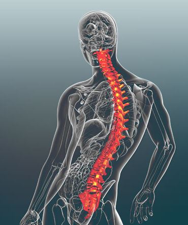
Adult degenerative scoliosis is a sideways curve of the spine that develops later in life as the discs and facet joints wear down. It is a different condition from the scoliosis diagnosed in teenagers, and it is treated differently.
Issada Thongtrangan, MD, treats adult degenerative scoliosis at Microspine in Scottsdale, Arizona, using endoscopic and minimally invasive techniques that preserve the muscles supporting the spine.
This page covers how the condition develops, how the decision to operate actually gets made, which patients qualify for an endoscopic approach, what recovery involves, and who should not have surgery.
How adult degenerative scoliosis differs from adolescent scoliosis
Adolescent idiopathic scoliosis develops during childhood or the teenage years, and the cause is unknown.
Adult degenerative scoliosis typically begins after the age of 50. It arises from the gradual breakdown of the spinal discs and facet joints. As one side of a disc wears down faster than the other, the spine begins to curve sideways. That curve is frequently accompanied by spinal stenosis, a narrowing of the spinal canal, and compression of the nerve roots.
That combination is why the leg symptoms are often more disabling than the curve itself.
What patients tell us
Adult scoliosis patients at Microspine are generally between 35 and 75 years old. This is how they describe the problem in their own words:
- “I can’t walk or stand longer than 5 to 10 minutes.”
- “I feel like I’m leaning sideways and pitching forward.”
- “My lower back muscles feel like they’re running a marathon 24/7 trying to keep me upright.”
- “My right or left leg goes completely numb and heavy if I stand up to cook dinner for more than ten minutes.”
The symptoms that bring patients in:
- Persistent lower back pain and stiffness
- Radicular pain, or sciatica, shooting down one or both legs
- Numbness, tingling or weakness in the legs, particularly when walking or standing for long periods, known as neurogenic claudication
- A visible forward or sideways lean in posture
Some people with a spinal curve have no symptoms at all. The size of the curve alone does not determine how much trouble it causes.
Types of scoliosis
Degenerative
Caused by thinning bone, disc wear and aging. This is the type most adults develop.
Idiopathic
No identifiable cause. Usually diagnosed in adolescence.
Neuromuscular
Secondary to a condition such as cerebral palsy or muscular dystrophy.
Congenital
Present from birth, caused by a vertebral abnormality.
Terms used to describe where and how a spine curves:
- Dextroscoliosis, a curve to the right
- Levoscoliosis, a curve to the left
- Thoracic scoliosis, a curve in the upper back
- Lumbar scoliosis, a curve in the lower back
- Thoracolumbar scoliosis, a curve spanning both
When surgery is the right answer
The degree of the curve is not what decides it. A patient with a 35 degree curve can live an active life with no pain, provided the curve is balanced.
Dr. T looks at three things instead:
- Walking tolerance. Whether walking distance is shrinking because of neurogenic claudication.
- Progression. Whether the curve is actively collapsing or worsening over time.
- Balance. Whether the patient is pitching forward or leaning noticeably to one side.
Surgery becomes the recommendation when conservative care has been exhausted and quality of life, independence or nerve function is actively deteriorating. Progressive neurological deficits, severe muscle fatigue and an inability to stay upright all move a patient toward surgery.
Treatment options, least invasive first
Conservative care
Patients with mild curves, intermittent discomfort or manageable symptoms are treated without surgery, using physical therapy, targeted injections and activity modification.
Endoscopic decompression
Endoscopic spine surgery uses a high-definition camera inserted through an incision a few millimeters wide. Through that channel Dr. T can see and decompress a pinched nerve directly, leaving the surrounding muscles and ligaments intact.
This suits a specific patient: sharp leg pain as the dominant complaint, minimal back pain, and no significant spinal imbalance. A patient who is pitching forward or leaning badly to one side will not be solved by decompression alone. Endoscopic technique also treats facet pain through targeted rhizotomy.
Minimally invasive fusion
When the spine needs stabilizing, specialized anterior or lateral approaches combined with percutaneous instrumentation allow the spine to be stabilized and realigned in selected patients, without a traditional long segmental fusion. MIS lateral lumbar interbody fusion is one of the approaches used.
When a longer fusion is necessary
Patients with significant instability or severe deformity may still need structural realignment. The choice depends on the severity of the curve, spinal balance, and whether the spine is unstable. Non-fusion options are considered first wherever they apply.
Why the ultra-minimally invasive approach matters:
- Smaller incisions and better cosmetic results
- Substantially less blood loss during surgery
- Shorter hospital stays, with many patients home the same day
- Faster recovery, and less reliance on narcotic pain medication afterward
What recovery actually looks like
- Hospital stay. 0 to 2 nights for minimally invasive and endoscopic procedures. Many patients leave the same day or the following morning. An overnight stay is typical after endoscopic or minimally invasive fusion.
- Walking. In the hallways within hours of surgery, with the care team.
- Driving. Around 2 to 3 weeks, once completely off narcotic pain medication and able to brake suddenly and safely.
- Desk or sedentary work. 2 to 6 weeks, depending on whether a fusion was performed, how many levels were treated, and your function before surgery.
- Active or physically demanding work. 8 to 12 weeks.
- Feeling like yourself again. Sharp leg pain commonly improves immediately after an endoscopic procedure. Muscle healing and stamina take roughly 3 to 6 months as the body adapts to improved alignment. Nerves heal slowly, and full nerve recovery can take up to 12 months.
These are typical ranges rather than guarantees. Recovery depends on the procedure performed, the number of levels treated, and your health going into surgery.
Who is not a candidate
Surgery is not the right tool for every patient, and saying so plainly is part of the evaluation. Dr. T advises against surgery for:
- Patients with mild, manageable symptoms who have not yet given non-surgical care a fair trial
- Patients expecting a long-standing, rigid, multi-level deformity to be fully reversed without the stabilization that correcting it requires
- Patients whose other medical conditions make the risks of anesthesia and spinal reconstruction outweigh the benefit
- Patients with untreated osteoporosis who would need a fusion
Being turned down for surgery does not end the conversation. Some patients are referred to physical therapy or a pain specialist. Others are treated to get them to candidacy, which can mean osteoporosis treatment, weight loss or stopping smoking.
Common questions about adult degenerative scoliosis
What is adult degenerative scoliosis, and how does it differ from adolescent scoliosis?
Adolescent idiopathic scoliosis develops during childhood or the teenage years with an unknown cause. Adult degenerative scoliosis typically begins after the age of 50 and arises from the gradual degeneration of spinal discs and facet joints. As one side of a disc wears down faster than the other, the spine begins to curve sideways, often accompanied by spinal stenosis and nerve root compression.
What are the common symptoms of adult degenerative scoliosis?
Some people with a spinal curve have no symptoms. Others notice persistent lower back pain and stiffness, radicular pain or sciatica shooting down one or both legs, numbness or tingling or weakness in the legs when walking or standing for long periods, and a noticeable forward or sideways lean in posture.
Who is a candidate for treatment and surgery?
Patients with mild curves, intermittent discomfort or manageable symptoms are treated non-surgically with physical therapy, targeted injections and activity modification. Surgery is considered for patients with persistent, debilitating pain, progressive neurological deficits, neurogenic claudication that severely limits walking distance, or a worsening deformity that has not responded to conservative treatment over several months. Endoscopic decompression suits patients whose symptoms come from focal nerve compression, a specific disc herniation or localized stenosis, particularly when sharp leg pain dominates and there is no significant spinal imbalance.
When is surgery recommended for adult degenerative scoliosis?
Surgery is considered when non-surgical treatment fails to relieve pain, or when a patient develops progressive neurological deficits, severe muscle fatigue, or an inability to stay upright that significantly affects daily quality of life.
How does endoscopic spine surgery apply to adult degenerative scoliosis?
Traditional open surgery for scoliosis involves large incisions, significant muscle stripping and extensive multi-level fusion. Many patients instead have focal nerve compression caused by a specific bone spur or disc herniation associated with their curve. Endoscopic spine surgery uses a high-definition camera through a millimeter-sized incision, allowing Dr. T to visualize and decompress the pinched nerve with precision tools while preserving the supporting muscles and ligaments.
Can endoscopic techniques replace traditional fusion for everyone with scoliosis?
No. The choice depends on the severity of the curve, spinal balance, and whether the spine is unstable. Patients with significant instability or severe deformity may still require structural realignment. In selected patients, endoscopic or minimally invasive fusion can take the place of an extensive long segmental fusion. Endoscopic techniques also address isolated nerve compression and treat facet pain through targeted rhizotomy, with minimal disruption to the body.
What are the benefits of the ultra-minimally invasive approach at Microspine?
Minimizing tissue trauma produces smaller incisions and better cosmetic results, significantly reduced blood loss during surgery, shorter hospital stays with many patients returning home the same day, faster overall recovery, and less reliance on narcotic pain medication afterward.