
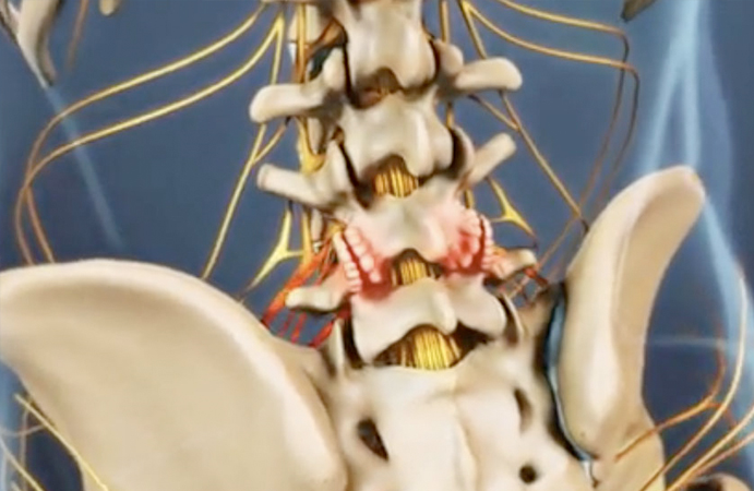
You may be a candidate for Endoscopic Rhizotomy
Facet joints are the small paired joints at the back of the spine that let you bend and twist. Like any joint, they wear out. When the cartilage thins and the joint becomes inflamed, the result is facet joint arthritis, also called facet syndrome or facet disease.
It is one of the most common causes of ongoing neck and lower back pain, and one of the most commonly misdiagnosed, because the pain it produces looks like several other things.
Dr. Issada Thongtrangan treats facet joint arthritis at Microspine in Scottsdale, Arizona. This page covers how the diagnosis is confirmed, what the treatment options are in order, and when surgery is and is not the answer.
What it feels like
- Aching pain in the lower back or neck, usually on one side, sometimes both
- Pain that is worse when leaning backward or twisting, and often better when sitting or leaning forward
- Stiffness that is worst first thing in the morning and after sitting still
- Pain that can refer into the buttock or the back of the thigh, and in the neck into the shoulder or the back of the head
- Pain that tends to build through the day rather than arriving suddenly
Facet pain usually stops above the knee. Pain traveling past the knee into the foot points to a compressed nerve rather than an arthritic joint, which is a different problem with a different treatment.
Causes
- Age-related wear of the cartilage in the joint, which is by far the most common
- Disc degeneration, which transfers load onto the facet joints behind it
- Previous injury to the joint from a fall, an accident or sport
- Repetitive strain from work or activity involving extension and rotation
- Spondylolisthesis, where a vertebra slips and changes how the joint is loaded
- Inflammatory arthritis affecting the spine
How facet pain is actually confirmed
This is the part that matters most, and the part patients are rarely told.
Facet joint arthritis shows up on imaging in most adults past a certain age, including plenty of people with no pain at all. A CT or MRI showing arthritic facet joints does not prove those joints are what hurts. Treating on imaging alone is how patients end up having a procedure that does not help.
Examination first. Pain on extension and rotation, tenderness over the joints, and a pattern of referred pain that matches the facet distribution.
Imaging to rule things out. X-ray, CT or MRI to see the joints and, just as importantly, to exclude nerve compression, instability or a disc problem producing similar symptoms.
A diagnostic block to prove it. A medial branch block numbs the specific small nerves carrying pain signals from the facet joint. If the pain goes away for the duration of the anesthetic, the joint is confirmed as the source. If it does not, the facet joint is not the problem and the plan changes.
Why the block comes before the treatment
The medial branch block is a test, not a cure. Its value is the answer it gives.
A rhizotomy targets exactly the nerves the block numbed. If numbing them did not relieve the pain, cutting their signal will not either.
Doing the diagnostic step properly is what separates a procedure that works from one that was never going to.
Treatment, least invasive first
Conservative care. Anti-inflammatory medication, physical therapy focused on core and postural strength, and activity modification. Most facet pain improves without anything further.
Facet joint injections. A steroid injected into or around the joint to reduce inflammation once the joint is confirmed as the source. Relief can last months and for some patients that is all that is needed.
Endoscopic rhizotomy. When the block confirms the joint and relief from injections is short-lived, the medial branch nerves can be interrupted directly. Dr. Thongtrangan uses an endoscopic approach, which means the nerve is seen under direct visualization through an incision a few millimeters wide rather than targeted blind.
MIS percutaneous facet fusion. When the problem is not only an inflamed joint but a mechanically unstable one, stabilizing the segment is the answer. This is the smallest version of that operation rather than an open fusion.
Why endoscopic rather than radiofrequency ablation
Radiofrequency ablation heats the medial branch nerve through a needle positioned by X-ray guidance. It is a reasonable option and it helps many patients. Its limitation is that the nerve is never actually seen, so success depends on the needle being in exactly the right place, and the nerve regenerates over time.
An endoscopic rhizotomy puts a camera on the nerve. Dr. Thongtrangan can see what he is treating, confirm it is the right structure, and treat it under direct vision. For patients who have had an ablation that worked briefly or not at all, that difference is usually the reason.
What recovery looks like
Injections and blocks. An outpatient appointment. Most patients drive themselves home and return to normal activity the next day.
Endoscopic rhizotomy. Day surgery. Home the same day, walking immediately, back to desk work within a few days and to physical work in two to four weeks.
Facet fusion. 0 to 2 nights in hospital. Walking in the hallways within hours. Driving at around 2 to 3 weeks once off narcotic pain medication. Desk work at 2 to 6 weeks, physically demanding work at 8 to 12 weeks.
These are typical ranges rather than guarantees. Recovery depends on the procedure, the number of levels treated, and your health going into it.
Who is not a candidate
Dr. Thongtrangan advises against a facet procedure for:
- Patients whose diagnostic block did not relieve the pain, because that result says the facet joint is not the source
- Patients whose pain is coming from a compressed nerve rather than the joint, which is a different problem treated differently
- Patients with mild, manageable symptoms who have not yet given conservative care a fair trial
- Patients whose other medical conditions make the risks outweigh the benefit
- Patients with untreated osteoporosis who would need a fusion
Being turned down does not end the conversation. Some patients are referred to physical therapy or a pain specialist. Others are treated to get them to candidacy.
Common questions about facet joint arthritis
What is facet joint arthritis?
Facet joints are the small paired joints at the back of the spine that allow bending and twisting. Facet joint arthritis, also called facet syndrome or facet disease, is the wearing down of the cartilage in those joints and the inflammation that follows. It is one of the most common causes of ongoing neck and lower back pain.
How do I know my pain is coming from the facet joints?
Imaging alone cannot tell you. Arthritic facet joints appear on scans in most adults past a certain age, including people with no pain at all. The pattern of symptoms points toward the facet joints, pain worse on leaning back and twisting, better sitting forward, usually stopping above the knee. Confirmation comes from a diagnostic medial branch block.
What is a medial branch block and why would I need one first?
The medial branch nerves carry pain signals from the facet joint to the spinal cord. A medial branch block numbs them temporarily. If your pain disappears while they are numb, the facet joint is confirmed as the source. If it does not, the facet joint is not the problem and the treatment plan changes. The block is a test rather than a cure, and its value is the answer it gives before anything irreversible is done.
What is an endoscopic rhizotomy?
An endoscopic rhizotomy interrupts the medial branch nerves so they stop carrying pain signals from an arthritic facet joint. It is performed through an incision a few millimeters wide using a high-definition camera, which means the nerve is seen directly rather than targeted by X-ray guidance alone. It is day surgery, and most patients go home the same day.
How is a rhizotomy different from radiofrequency ablation?
Radiofrequency ablation heats the nerve through a needle positioned using X-ray guidance, so the nerve itself is never seen. An endoscopic rhizotomy places a camera on the nerve, so the structure is confirmed visually and treated under direct vision. Patients who have had an ablation that gave brief or no relief are often candidates for the endoscopic approach for exactly that reason.
Do I need a fusion for facet joint arthritis?
Usually not. An inflamed, arthritic joint and a mechanically unstable one are different problems. Where the joint is painful but stable, treating the pain source directly through injections or an endoscopic rhizotomy addresses what is actually wrong. Fusion is the right answer when the segment is genuinely unstable, and it is the smaller percutaneous version of that operation rather than an open fusion wherever possible.
What does recovery involve?
Injections and diagnostic blocks are outpatient appointments, with most patients back to normal activity the next day. An endoscopic rhizotomy is day surgery, home the same day, walking immediately, desk work within a few days and physical work in two to four weeks. A facet fusion involves 0 to 2 nights in hospital, driving at around 2 to 3 weeks, desk work at 2 to 6 weeks and physical work at 8 to 12 weeks. These are typical ranges rather than guarantees.